|
 Magnetic resonance imaging (MRI) uses radiofrequency waves and a strong magnetic field rather than x-rays to provide remarkably clear and detailed pictures of internal organs and tissues. The technique has proven very valuable for the diagnosis of a broad range of pathologic conditions in all parts of the body including cancer, heart and vascular disease, stroke, and joint and musculoskeletal disorders. MRI requires specialized equipment and expertise and allows evaluation of some body structures that may not be as visible with other imaging methods. Magnetic resonance imaging (MRI) uses radiofrequency waves and a strong magnetic field rather than x-rays to provide remarkably clear and detailed pictures of internal organs and tissues. The technique has proven very valuable for the diagnosis of a broad range of pathologic conditions in all parts of the body including cancer, heart and vascular disease, stroke, and joint and musculoskeletal disorders. MRI requires specialized equipment and expertise and allows evaluation of some body structures that may not be as visible with other imaging methods.
What are some common uses of the MRI procedure?
 MRI scans are particularly suited to imaging of non-bony parts and soft tissues within the body. The brain, spinal cord, nerves, muscles, ligaments, and tendons are all seen on MRI scans more clearly than on other scans such as x-rays or CT scans. AN MRI can differentiate between gray and white matter.
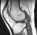
Because MRI can give such clear pictures of soft-tissue structures near and around bones, it is the most sensitive exam for spinal and joint problems. MRI is widely used to diagnose sports-related injuries, especially those affecting the knee, shoulder, hip, elbow and wrist. The images allow the physician to see even very small tears and injuries to ligaments and muscles.
MRI scans are particularly suited to imaging of non-bony parts and soft tissues within the body. The brain, spinal cord, nerves, muscles, ligaments, and tendons are all seen on MRI scans more clearly than on other scans such as x-rays or CT scans. AN MRI can differentiate between gray and white matter.
Because MRI can give such clear pictures of soft-tissue structures near and around bones, it is the most sensitive exam for spinal and joint problems. MRI is widely used to diagnose sports-related injuries, especially those affecting the knee, shoulder, hip, elbow and wrist. The images allow the physician to see even very small tears and injuries to ligaments and muscles.
In addition, MRI of the heart, aorta, coronary arteries and blood vessels is a fast, noninvasive tool for diagnosing coronary artery disease and heart problems. Physicians can examine the size and thickness of the chambers of the heart and determine the extent of damage caused by a heart attack or progressive heart disease.
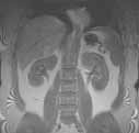
Organs of the chest and abdomen-including the lungs, liver, kidney, spleen, pancreas and abdominal vessels-can also be examined in high detail with MRI, enabling the diagnosis and evaluation of tumors and functional disorders. MRI is growing in popularity as an alternative to traditional x-ray mammography in the early diagnosis of breast cancer.
Because an MRI does not use x-rays or other radiation, it is commonly used when frequent imaging may be required for diagnosis or ongoing monitoring, especially in vulnerable areas like the brain. Specialized types of MRIs may also be called for, such as a functional MRI that is used to observe brain structures and determine which areas of the brain "activate" during various cognitive tasks. This can help advance understanding of brain organization.
How should I prepare for the procedure?
Because the strong magnetic field used for MRI will pull on any ferromagnetic metal object implanted in the body, MRI staff will ask whether you have a prosthetic hip, heart pacemaker (or artificial heart valve), implanted port, infusion catheter (brand names Port-o-cath, Infusaport, Lifeport), intrauterine device (IUD), or any metal plates, pins, screws or surgical staples in your body. In most cases surgical staples, plates, pins and screws pose no risk during MRI if they have been in place for more than four to six weeks. Tattoos and permanent eyeliner may also create a problem. You will be asked if you have ever had a bullet or shrapnel in your body or ever worked with metal. If there is any question of metal fragments, you may be asked to have an x-ray that will detect any such metal objects. Tooth fillings usually are not affected by the magnetic field but they may distort images of the facial area or brain, so the radiologist should be aware of them. The same is true of braces, which may make it hard to "tune" the MRI unit to your body. You will be asked to remove anything that might degrade MRI images of the head, including hairpins, jewelry, eyeglasses, hearing aids and any removable dental work.
The radiologist or technologist may ask about drug allergies and whether head surgery has been done in the past. If you might be pregnant, this should be mentioned. Some patients who undergo MRI in an enclosed unit may feel confined or claustrophobic. If you are not easily reassured, a sedative may be administered. Roughly one in 20 patients will require medication to reduce the anxiety associated with claustrophobia.
What does the MRI equipment look like?
The conventional MRI unit is a closed cylindrical magnet in which the patient must lie totally still for several seconds at a time and consequently may feel "closed-in" or truly claustrophobic. However, new "patient-friendly" designs are rapidly coming into routine use.
The "short-bore" systems are wider and shorter and do not fully enclose the patient. Some newer units are open on all sides, however the image quality may vary. Examples of the radiography equipment that may be used are shown at the top of this page.
How does the procedure work?
MRI is a unique imaging method because, unlike the usual radiographs (x-rays), radioisotope studies or even Computed Tomography (CT) scanning, it does not rely on ionizing radiation. Instead radiofrequency waves are directed at protons, the nuclei of hydrogen atoms, in a strong magnetic field. The protons are first "excited" and then "relaxed," emitting radio signals that can be computer-processed to form an image. In the body, protons are most abundant in the hydrogen atoms of water-the "H" of H2O -so that an MR image shows differences in the water content and distribution in various body tissues. Even different types of tissue within the same organ, such as the gray and white matter of the brain, can easily be distinguished. Typically an MRI examination consists of two to six imaging sequences, each lasting two to 15 minutes. Each sequence has its own degree of contrast and shows a cross-section of the body in one of several planes (right to left, front to back, upper to lower).
How is the procedure performed?
 The patient is placed on a sliding table and positioned comfortably for the MRI examination. Then the radiologist and technologist leave the room and the individual MRI sequences are performed. The patient is able to communicate with the radiologist or technologist at any time using an intercom. Also, many MRI centers allow a friend or, if a child is being examined, a parent to stay in the room. The patient is placed on a sliding table and positioned comfortably for the MRI examination. Then the radiologist and technologist leave the room and the individual MRI sequences are performed. The patient is able to communicate with the radiologist or technologist at any time using an intercom. Also, many MRI centers allow a friend or, if a child is being examined, a parent to stay in the room.
Depending on how many images are needed, the exam will generally take 15 to 45 minutes, although a very detailed study may take longer. You will be asked not to move during the actual imaging process, but between sequences some movement is allowed. Patients are generally required to remain still for only a few seconds to a few minutes at a time.
Depending on the part of the body being examined, a contrast material (usually gadolinium) may be used to enhance the visibility of certain tissues or blood vessels. A small needle connected to an intravenous line is placed in an arm or hand vein. A saline solution will drip through the intravenous line to prevent clotting until the contrast material is injected about two-thirds of the way through the exam.
When the exam is over the patient is asked to wait until the images are examined to determine if more images are needed. A radiologist experienced in MRI will analyze the images and send a report with his or her interpretation to the patient's personal physician. This should take only a few days or less.
What will I experience during the MRI procedure?
MRI causes no pain but some patients can find it uncomfortable to remain still during the examination. Others experience a sense of being "closed in," though the more open construction of newer MRI systems has done much to reduce that reaction. You may notice a warm feeling in the area under examination; this is normal but if it bothers you the radiologist or technologist should be notified.
If a contrast injection is needed, there may be discomfort at the injection site and you may have a cool sensation at the site during the injection. Most bothersome to many patients are the loud tapping or knocking noises heard at certain phases of imaging. Ear plugs may help.
Who interprets the results and how do I get them?
A radiologist, who is a physician experienced in MRI and other radiology examinations, will analyze the images and send a signed report with his or her interpretation to the patient's personal physician. The patient receives MRI results from the referring physician who ordered the test. New technology also allows for distribution of diagnostic reports and referral images over the Internet at many facilities.
What are the benefits vs. risks?
Benefits
- Images of the soft-tissue structures of the body-such as the heart, lungs, liver and other organs-are clearer and more detailed than with other imaging methods.
- MRI can help physicians evaluate the function as well as the structure of many organs.
- The detail makes MRI an invaluable tool in early diagnosis and evaluation of tumors.
- MRI contrast material is less likely to produce an allergic reaction than the iodine-based materials used for conventional x-rays and CT scanning.
- MRI enables the detection of abnormalities that might be obscured by bone with other imaging methods.
- MRI provides a fast, noninvasive alternative to x-ray angiography for diagnosing problems of the heart and cardiovascular system.
- Exposure to radiation is avoided.
Risks
- An undetected metal implant may be affected by the strong magnetic field.
- MRI is generally avoided in the first 12 weeks of pregnancy. Doctors usually use other methods of imaging, such as ultrasound, on pregnant women unless there is a strong medical reason to use MRI.
What are the limitations of MRI of the Body?
Bone is better imaged by conventional x-rays in some cases and CT is preferred for patients with severe bleeding. MRI may not always distinguish between tumor tissue and edema fluid and does not detect calcium when this is present within a tumor. In most cases the examination is safe for patients with metal implants, with the exception of a few types of implants, so patients should inform the technician of an implant prior to the test. The examination must be used cautiously in early pregnancy. MRI typically costs more than CT scanning.
|